The Physics of Fragmentation

There’s a lot of confusion about how DNA and chromatin are fragmented using sonication in preparing samples for downstream assays such as PCR or sequencing. We’re here to set the record straight, and to show you the fun side of the Physics of Fragmentation.

When an ultrasound wave propagates through a fluid, the associated energy is quite low and diffuse. Fragmentation of DNA or chromatin is mediated by bubbles. Bubbles concentrate the diffuse energy into very small spatial and temporal scales, allowing the bubbles to do work on their surroundings. There are several bubble-mediated mechanisms that can cause fragmentation. These include mechanical impact (or water hammer), shock waves, microstreaming, heat and radicals. Let’s look at them in more detail.

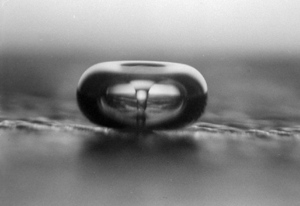
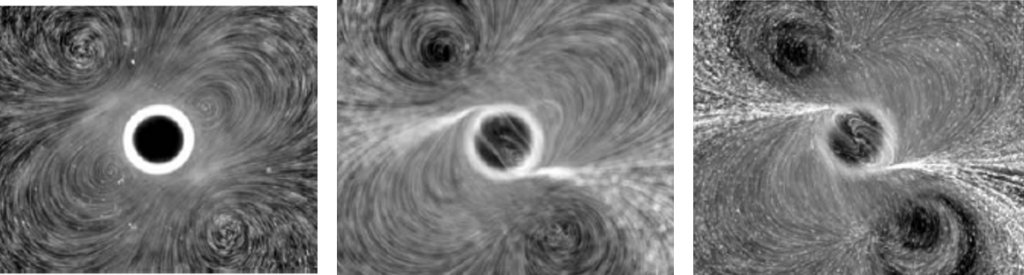
Water hammer: One of the most widely circulated mechanisms in the scientific literature involves direct mechanical impact associated with cavitation collapse. The mechanism is best illustrated by looking at a bubble collapsing against a solid surface. Figure 1a shows a high-speed video sequence of such a bubble. The jet that’s formed can be very powerful. It acts as a water hammer and can pit even hard objects like boat propellers. The jet velocity can be hundreds of meters/second, leading to impact pressures of hundreds of MPa. Figure 1b shows a closeup of the jet and a video of the dynamics. It’s important to note that jets form in the absence of surfaces too; all that’s required is something to cause an asymmetry in the fluid flow around the bubble, such as another nearby bubble.

Figure 1a. Bubble collapse near a surface. Bubble collapse leads to a fluid jet. Citation: Werner Lauterborn and Claus-Dieter Ohl, “The Peculiar Dynamics of Cavitation Bubbles,” Applied Scientific Research, Vol. 58, pp 63–76 (1998).

Figure 1b. Liquid jet through a bubble. Image and movie courtesy of Lawrence Crum, Ph.D.


Shock waves: Another mechanism for shearing is shock waves. When a bubble collapses symmetrically, the implosion can lead to the emission of an acoustic shock wave. The shock front is a place for rapid changes in stress, temperature, and density. Figure 2 shows a high speed video movie of a bubble collapse leading to the emission of a shock wave.

Figure 2. Shock wave emission from a spherical bubble collapse. Video courtesy of Thomas J. Matula, Ph.D.

Sonoluminescence: The shock wave emission video above also brings up a question about the bubble interior. If the collapse is so violent it can generate a shock wave, what are the conditions inside the bubble? Imagine a collapsing bubble. Initially, there’s plenty of time for heat to equilibrate, and the process is considered isothermal. But during the final stages, the bubble may collapse adiabatically. Adiabatic heating of the bubble interior – specifically the center of the bubble – can lead to very high temperatures (hotter than the surface of the sun!), chemical dissociation of molecular bonds, ionization and radical formation, and even the emission of light, called sonoluminescence. Figure 3a shows the dynamics of how a bubble can grow and collapse symmetrically to generate a light flash. Figure 3b shows the light emission. In reality, the light comes from the very center of the bubble, lasting less than a nanosecond. The video averages out the flashes and it looks like it’s continuously emitting, but that’s not the case.
Figure 3. Sonoluminescence bubble. (a) Bubble dynamics. Citations: Thomas J. Matula, “Inertial Cavitation and sonoluminescence,” Phil. Trans. R. Soc. Lond., 357, 225-249 (1999). Thomas J. Matula, “Single-bubble sonoluminescence in microgravity,” Ultrasonics 38, 559-565 (2000). (b) Standard video of light emission. Video courtesy of Thomas J. Matula, Ph.D.
Microstreaming: Cavitation induces microstreaming which generates large shear stresses on nearby objects. This phenomenon is probably the most important mechanism for DNA fragmentation. An example image and movie is shown in figure 4. At low pressures, microstreaming can help remove particles from surfaces, while at higher pressures, the microstreaming can be intense.
Figure 4. Shear stresses induced on particles near an oscillating bubble. (a) Citation: Paul Tho, Richard Mannasseh, and Andrew Ooi, “Cavitation microstreaming patterns in single and multiple bubble systems,” Journal of Fluid Mechanics; Vol. 576, pp 191-233 (2007). (b) Video courtesy of Richard Mannasseh, Ph.D.
Frequency effects: The mechanisms described above can occur at frequencies ranging from 20 kHz (or lower) all the way up into the MHz range. Also, all sonication instruments generate cavitation. The most basic lab instrument is the ultrasonic horn (also called a “cell disruptor,” “homogenizer,” “Sonotrode,” etc.). It operates at the low end of the frequency spectrum, around 20-40 kHz or so. The low end is determined by the desire to stay above the threshold for hearing. Hence, you won’t see any instruments working below 20 kHz. Even those operating at 20-40 kHz can generate a significant amount of audible noise, because of vibrations induced in other components. The high end of operation is limited by the ability of cavitation to do work. When a bubble grows, it absorbs potential energy, and that energy is converted into kinetic energy during collapse, which is then used to do work. Making a bubble grow for a longer period of time (=low frequency) would impart more energy into the system, while making a bubble oscillate more frequently (= high frequency) allows the bubble to do more work per unit time.

Sample Heating: All bubbles absorb energy from the acoustic wave, and some of that energy is dissipated as heat. That’s the physics, and there’s no getting around it. Heating is controlled by cooling the samples and optimizing acoustic pulse parameters.